
X射線探測
X射線檢測器中使用的閃爍體材料需要高X射線吸收效率和光輸出,低余輝,透明性以及與光電轉(zhuǎn)換器匹配的光譜響應(yīng)。不同閃爍體的性能差異很大。為了與特定的CCD配合使用,我們應(yīng)該綜合考慮發(fā)光效率,光譜響應(yīng)和其他特性,并選擇合適的閃爍體材料。
閃爍體中X射線輻射的平均吸收深度取決于光子能量和材料。材料透明度的優(yōu)勢隨成像板的厚度而降低。如果閃爍體更薄,則平均吸收深度會更低,并且由于閃爍光子的橫向擴展較少而導(dǎo)致的圖像模糊較少,因此生成的圖像會更清晰。因此,成像板越薄,圖像中獲得的分辨率越好。另一方面,隨著閃爍體的厚度,檢測效率降低。
基于CCD的X射線探測器由閃爍體,光耦合器和CCD陣列組成。 它覆蓋了CCD線性陣列上的閃爍體薄層。 閃爍體將X射線轉(zhuǎn)換為對CCD敏感的可見光,然后將CCD轉(zhuǎn)換為反映X射線在空間中分布的電信號。
應(yīng)用于X射線探測的閃爍晶體
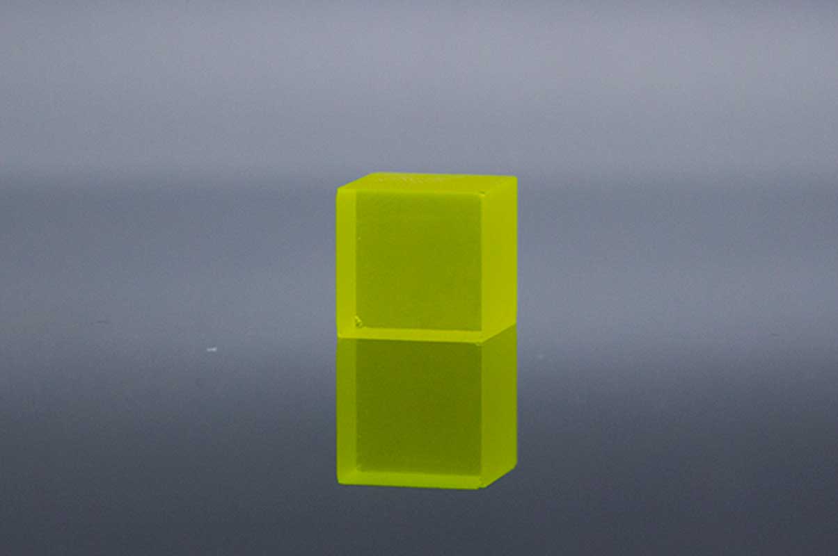
Ce:YAG
| 波長(最大發(fā)射)(nm) | 550 |
| 衰減時間(ns) | 70 |
| 發(fā)光量(光子/ keV) | 35 |
| 折光率 | 1.82@550nm |
| 輻射長度(cm) | 3.5 |
Ce:YAG是一種快速閃爍體,具有優(yōu)異的機械和化學(xué)耐受性,其衰減時間約為70 ns,這使得高計數(shù)率成為可能,并且量子產(chǎn)率估計為 介于40至50 ph / keV之間,盡管光輸出較小,但它的非吸濕性和較短的衰減時間與標(biāo)準(zhǔn)CsI:Tl閃爍體相比仍具有優(yōu)勢,最大發(fā)射波長(約550 nm)非常適合 PMT模塊的光陰極的靈敏度。實驗證明,YAG:Ce和LuAG:Ce屏幕適合于高空間分辨率的成像。所提出的成像系統(tǒng)的分辨率約為1微米。
參考文獻
[1] An X-ray counting system based on YAG Ce scintillator
[2] Thin YAG:Ce and LuAG:Ce single crystal imaging plates used for high spatial resolution in X-ray imaging systems
[3] Similarity of trap state and thermoluminescence processes of Ce YAG for X-ray and UV irradiation
[4] High-resolution imaging of biological and other objects with an X-ray digital camera
[5] High-resolution application of YAG Ce and LuAG Ce imaging detectors with a CCD X-ray camera
Ce:YAP
| 波長(最大發(fā)射)(nm) | 370 |
| 衰減時間(ns) | 28 |
| 發(fā)光量(光子/ keV) | 25 |
| 相對于Nal(Tl)的光輸出(%) | 60-70 |
| 折光率 | 1.95@370nm |
Ce:YAP晶體是出色閃爍體的特性包括:(i)30 ns的快速衰減時間,(ii)7.4 g / cm3的高密度,和(iii)約30%的高光產(chǎn)率 在NaI(Tl)閃爍體中 原鋁酸釔(YAlO3:Ce or YAP:Ce)是快速發(fā)射閃爍體,主要用于PET和動物PET檢測器中
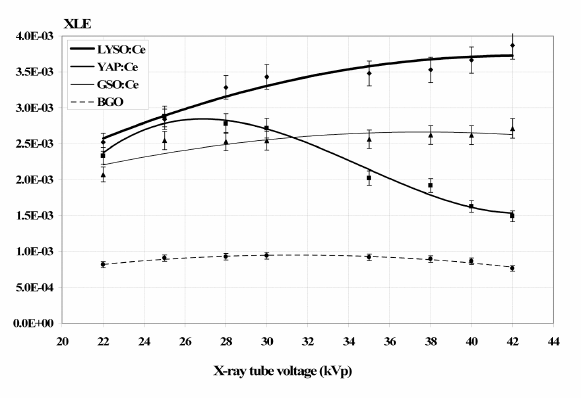
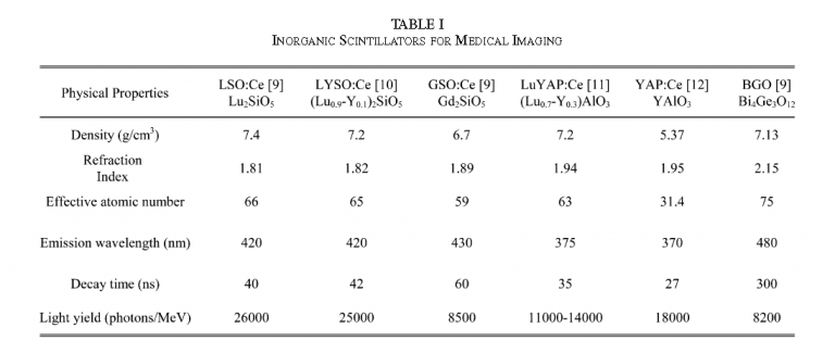
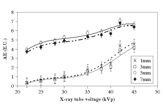
The x-ray luminescence efficiency (XLE) of Ce:LYSO, Ce:YAP, Ce:GSO and BGO as determined by the experimental data for x-ray tube voltages between 22–42kVp(mammography).Points: measured data, line: fitted curve.
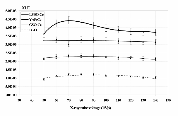
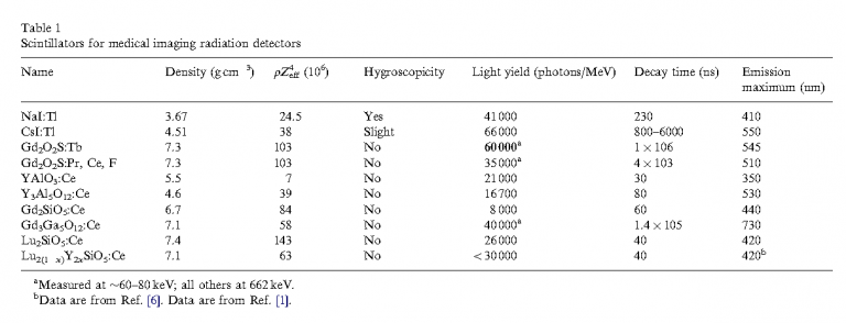
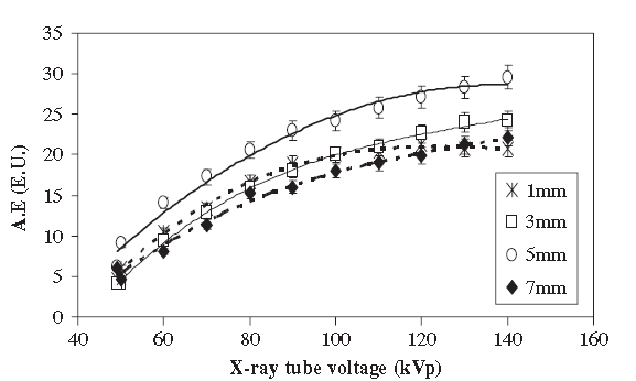
The x-ray luminescence efficieny (XLE) of Ce:LYSO, Ce:YAP, Ce:GSO and BGO as determined by the experimental data for x-ray tube voltages between 50–140 kVp (general radiography).Points: measured data, line: fitted curve.
參考文獻
[1] A YAP(Ce) imager operated in high energy X-ray region
[2] Comparative evaluation of single crystal scintillators under x-ray imaging conditions
[3] Comparative Investigation of Ce Doped Scintillators in a Wide Range of Photon Energies Covering X-ray CT, Nuclear Medicine and Megavoltage Radiation Therapy Portal Imaging Applications
[4] ISPA tubes with scintillating YAP Ce windows X- and γ- ray imaging
[5] High-resolution application of YAG Ce and LuAG Ce imaging detectors with a CCD X-ray cameraMcps-range photon-counting X-ray computed tomography system utilizing an oscillating linear-YAP(Ce) photon detector
[6] Properties of a YAP Ce detector for high-energy X-ray counting experiments
Ce:LYSO

| 波長(最大發(fā)射)(nm) | 410 |
| 衰減時間(ns) | 40 |
| 發(fā)光量(光子/ keV) | 25 |
| 相對于Nal(Tl)的光輸出(%) | 75 |
| 折光率 | 1.82@410nm |
基于镥-釔的閃爍體,例如LYSO:Ce,具有很高的有效原子序數(shù),是非吸濕的,快速發(fā)射的材料,并且有望用于正電子發(fā)射成像儀。
Ce: LYSO是混合的LSO / YSO(5-10%)非吸濕性晶體,具有高密度(7.1 g / cm3),高光輸出(30000 ph / MeV),良好的能量分辨率(10%)和短衰減 時間(40 ns)。 盡管Ce: LYSO和Ce: LSO表現(xiàn)出相似的行為,但就衰減方案而言,在低能(35 kV)X射線激發(fā)下,Ce: LYSO的光產(chǎn)率似乎比LSO高約20%。
參考文獻
[1] A comparative study of the luminescence properties of LYSO Ce, LSO Ce, GSO Ce and BGO single crystal scintillators for use in medical X-ray imaging
[2] Comparative evaluation of single crystal scintillators under x-ray imaging conditions
[3] Comparative Investigation of Ce Doped Scintillators in a Wide Range of Photon Energies Covering X-ray CT, Nuclear Medicine and Megavoltage Radiation Therapy Portal Imaging Applications
[4] Evaluation of the light emission efficiency of LYSO Ce scintillator under X-ray excitation for possible applications in medical imaging
[5] Improving Ce3+ doped scintillating materials for medical imaging applications
[6] Investigation of luminescence emission properties of LYSO Ce and LuYAP Ce single crystal scintillators under x-ray exposure for use in medical imaging
[7] Luminescence Properties of LYSO ce and GSO Ce Single Crystal Scintillators Under X-Ray Excitation for Use in Medical Imaging Systems
[8] Scintillator efficiency study with MeV x-rays
Ce:GAGG
| 波長(最大發(fā)射)(nm) | 520 |
| 波長范圍(nm) | 475-800 |
| 衰減時間(ns) | 90 |
| 發(fā)光量(光子/ keV) | 50 |
| 折光率 | 1.95@540nm |
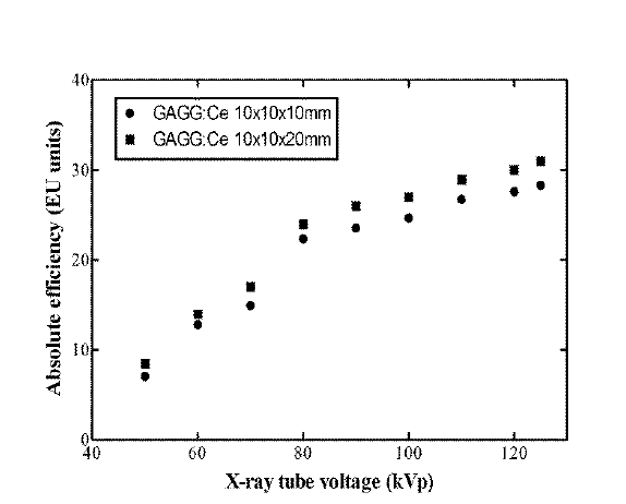
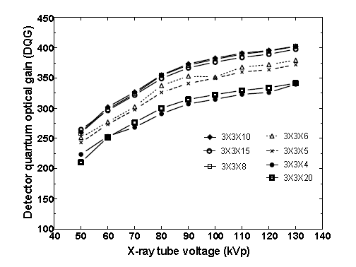
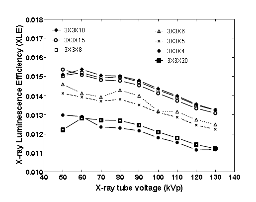
Ce:GAGG (Ce:Gd3Al2Ga3O12) 閃爍晶體,具有高密度(6.69 g/cm-3),快速閃爍響應(yīng)(?90 ns)和高光產(chǎn)率(?46000 ph / MeV) )。 與目前許多高效的閃爍體相比,GAGG:Ce具有非吸濕性(如LaBr3:Ce,LaCl3:Ce,CsI和NaI:Tl晶體),并且不具有天然放射性(如基于based的材料)。 GAGG:Ce發(fā)出520nm的光子,有效原子序數(shù)等于54.4。
參考文獻
[1] X-ray Luminescence Efficiency of GAGG Ce Single Crystal Scintillators for use in Tomographic Medical Imaging System
[2] Light Emission Efficiency of GAGG Ce Single Crystal Under X-ray Radiographic Conditions
Ce:LuAG
| 波長(最大發(fā)射)(nm) | 535 |
| 波長范圍(nm) | 475-800 |
| 衰減時間(ns) | 70 |
| 發(fā)光量(光子/ keV) | 25 |
| 折光率 | 1.84@633nm |
高分辨率成像系統(tǒng)是由高靈敏度數(shù)字CCD攝像機和光學(xué)系統(tǒng)與薄閃爍成像屏組成。屏幕可以包括LuAG:Ce無機閃爍體。
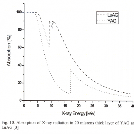
[參考文獻1]比較了兩種屏幕的發(fā)光效率(每keV可見光子數(shù))和空間分辨率。在第一個實驗中,CCD檢測到的光在CCD中心200μ200像素的平方ROI中平均。這個Ce:YAG發(fā)射值17507(每秒CCD像素中的電子數(shù))和Ce:LuAG給出值26452,大約是Ce:YAG。這個Ce:LuAG單曲晶體比釔鋁石榴石(密度:6.73–4.57 g/cm3)并且X射線被LuAG吸收得更強(在1到40 keV范圍內(nèi)吸收的X射線輻射(光子)是X射線輻射衰減系數(shù)的1.7倍)。這個Ce:LuAG屏幕具有比Ce:YAG屏使圖像的信噪比更好地應(yīng)用于成像系統(tǒng)中。
[參考文獻2]使用Ce:LuAG 20 mm屏幕拍攝圖像。CCD相機的有效像素尺寸為0.74mm。X射線微聚焦源的工作電壓為40kV / 2mA。圖像采集時間為5s,對25個圖像樣本進行平均。在從源發(fā)出的光子通量轉(zhuǎn)換后,Ce:LuAG產(chǎn)生的光的測量強度約為Ce:YAG值的1.51倍。用CCD檢測光,并在200×200像素的平方ROI中求平均值。與Ce:YAG(密度:6.73–4.57g / cm3)相比,Ce:LuAG單晶密度更高,LuAG對X射線的吸收更強(X射線的平均吸收量是1.7倍)。1至40keV之間的范圍)
參考文獻
[1] High-resolution application of YAG Ce and LuAG Ce imaging detectors with a CCD X-ray camera
[2] High-resolution imaging of biological and other objects with an X-ray digital camera
[3] Thin YAG Ce and LuAG Ce single crystal imaging plates used for high spatial resolution in X-ray imaging systems
Tl:CsI
| 波長(最大發(fā)射)(nm) | 410 |
| 衰減時間(ns) | 40 |
| 發(fā)光量(光子/ keV) | 25 |
| 相對于Nal(Tl)的光輸出(%) | 75 |
| 折光率 | 1.82@410nm |
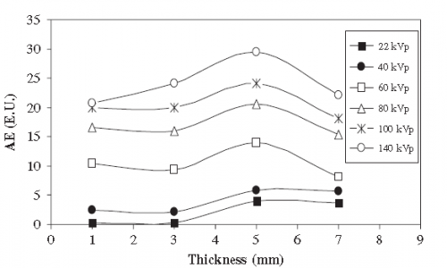
CsI(Tl)的最重要特征是其高光產(chǎn)率(≥104光子/ MeV),并且其發(fā)射光譜在約550 nm處具有最大值,與非晶和結(jié)晶硅光電二極管完全兼容。研究了CsI:Tl單晶閃爍體的發(fā)光效率與X射線乳腺攝影和一般X射線成像所用能量范圍內(nèi)的晶體厚度和X射線管電壓的關(guān)系。
[參考文獻1]絕對發(fā)光效率,這是在X射線成像(40–140 kV)和乳房X線照片成像(22–49 kV)中使用的各種X射線能量中通過實驗確定的。使用以下設(shè)備通過X射線對晶體進行輻照:(i)具有鎢陽極靶和2mm Al濾光片的飛利浦X射線裝置和(ii)帶有鉬陽極靶和鉬濾光片的通用電子Senographer DMR X射線乳腺攝影裝置,使用前面描述的實驗裝置,對晶體進行X射線輻照。
[參考文獻2]為了滿足這些需求,我們使用結(jié)構(gòu)化的CsI(TI)閃爍體與快速幀1K x 1K CCD耦合,開發(fā)了一種原型快速X射線成像系統(tǒng)。該系統(tǒng)已成功用于以12位動態(tài)范圍以每秒1000幀(fps)的速率捕獲1024 x 64像素x射線圖像。該系統(tǒng)超越了通常以30 fps的速率運行的當(dāng)前高速X射線成像系統(tǒng)的功能。CsI(TI)(59,000個光子I MeV)的高x射線轉(zhuǎn)換效率使這些屏幕成為當(dāng)前x射線衍射應(yīng)用的一個極好的選擇,在x射線衍射中,某些重要的衍射峰往往很弱,需要具有高信噪比的成像屏幕才能正確識別。
參考文獻
[1] A systematic study of the performance of the CsI Tl single-crystal scintillator under X-ray excitation
[2] high-speed-xray-imaging-camera-using-structured-csitl-scintillator
[3] Scintillator efficiency study with MeV x-rays
[4] Structured CsI(Tl) scintillators for X-ray imaging applications
[5] Validation of columnar CsI x-ray detector responses obtained with hybridr , a CPU-GPU Monte Carlo code for coupled x-ray, electron, and optical transport
CdWO4
| 波長(最大發(fā)射)(nm) | 490 |
| 衰減時間(ns) | 14000 |
| 發(fā)光量(光子/ keV) | 12-15 |
| 相對于Nal(Tl)的光輸出(%) | 50 |
| 折光率 | 2.2-2.3 |
鎢酸鎘(CdWO4,CWO)晶體是此類應(yīng)用的候選者之一,因為它具有較高的有效Z值,高密度,比BGO更高的光產(chǎn)率以及非常低的余輝。CWO晶體的輻射硬度高達105 rad。由于這種優(yōu)勢,CWO閃爍體被廣泛用于X射線CT中。
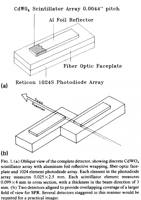
[參考文獻1]描述了一種用于X射線成像的高分辨率X射線檢測器陣列,它由CdWO4閃爍體的離散陣列和帶有光纖面板的Reticon 1024S光電二極管陣列組成。它開發(fā)了一種非常高分辨率的離散閃爍體陣列,用于扇形束X射線成像系統(tǒng)。
參考文獻
[1] High-resolution digital x-ray detector utilizing a discrete array of CdWO4 scintillators and a self-scanned photodiode array
[2] Large Size CdWO4 Crystal for Energetic X- and γ Ray Detection
[3] X-RAY STUDY OF CHARACTERIZATION OF THE PbWO4 AND CdWO4 SINGLE CRYSTALS
GOS
| 波長(最大發(fā)射)(nm) | 510 |
| 衰減時間(us) | 5.5 |
| 相對光輸出(%) | 80 |
| 余輝(%) | <0.01 |
| 發(fā)光強度(keV) | 27.5 |
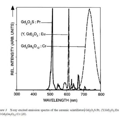
| GOS(Gd2O2S) | Density (g/cm3) | Wavelength of max emission (nm) | Decay constant (ns) |
| Ref[1] | 7.34 | 510 | 5.5μs |
參考文獻